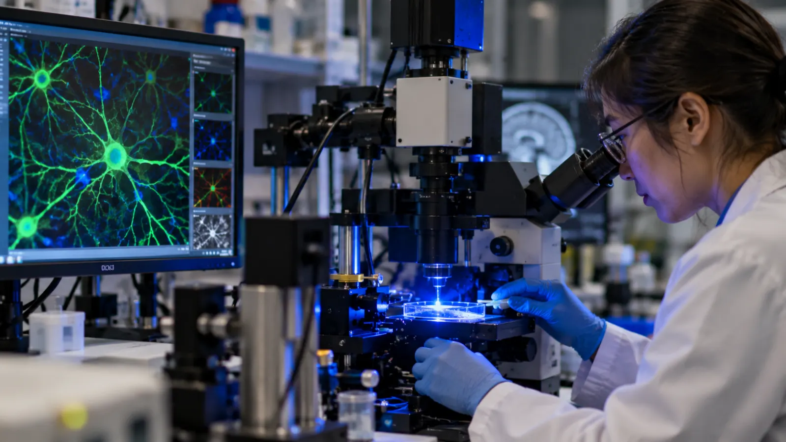
Nobel Prize in Medicine 2026: How Light Can Control Brain Cells
The 2026 Nobel Prize in Physiology or Medicine has been awarded jointly to Karl Deisseroth, Peter Hegemann and Georg Nagel for discoveries involving light-gated ion channels and the development of optogenetics, a technique that allows scientists to control selected nerve cells using light. Their work has transformed neuroscience by giving researchers a way to move beyond simply observing which parts of the brain become active and instead test what particular groups of neurons actually do.
The prize, announced on October 5, carries 12 million Swedish kronor, to be shared by the three scientists. Deisseroth is associated with Stanford University and the Howard Hughes Medical Institute in the United States, Hegemann works at Humboldt University in Berlin, and Nagel is based at the University of Würzburg in Germany. Their discoveries developed over decades, beginning with an apparently simple biological question about how microscopic algae respond to light and eventually producing one of modern neuroscience’s most powerful experimental tools.
What is optogenetics?
Optogenetics combines genetics and light to control the activity of selected cells. It is used most famously in neuroscience, where researchers can make specific neurons sensitive to light and then activate or silence them with extraordinary precision.
To understand why this matters, consider how the brain works. The human brain contains billions of neurons communicating through electrical and chemical signals. Different networks of neurons contribute to movement, memory, fear, reward, sleep, vision and many other processes. Scientists have long been able to measure brain activity and identify regions associated with particular behaviours, but observing activity does not necessarily prove that those cells caused the behaviour.
Optogenetics changed that. Researchers can introduce genes encoding light-sensitive proteins into carefully selected neurons. When those proteins are exposed to particular wavelengths of light, they allow electrically charged ions to move across the cell membrane. This changes the electrical state of the neuron and can cause it to become active or, depending on the protein used, reduce its activity.
In simple terms, researchers gained something resembling a highly precise light switch for nerve cells.
If activating a particular group of neurons causes an animal to perform a behaviour, scientists gain stronger evidence that those cells participate directly in producing that behaviour. If switching the neurons off prevents the behaviour, the causal relationship becomes even clearer.
This ability to manipulate identified brain circuits in real time has fundamentally changed how researchers investigate the nervous system.
The discovery began with a tiny green alga
The origins of the Nobel-winning research are surprisingly far removed from human medicine.
Peter Hegemann was interested in Chlamydomonas, a microscopic single-celled green alga capable of moving towards light. For the organism to do this, it needs a biological mechanism capable of detecting light and rapidly converting that information into a cellular response.
Working on these light-sensitive mechanisms, Hegemann and Georg Nagel helped identify and characterise a protein known as channelrhodopsin.
Channelrhodopsins sit in the cell membrane and respond directly to light. When illuminated with light of the appropriate wavelength, the protein changes shape and opens a channel. Charged particles, known as ions, can then flow through the opening.
Because electrical activity inside cells depends heavily on the movement of ions across membranes, this discovery provided something researchers had long wanted: a biological mechanism that could convert light directly into an electrical cellular signal.
An especially important form, channelrhodopsin-2, responds to blue light. Researchers discovered that the protein could function even when introduced into cells very different from the algae in which it naturally occurs.
That opened the door to a much more ambitious idea.
Could a light-sensitive protein from algae be placed inside a neuron and used to control the neuron with light?
How Karl Deisseroth helped turn the discovery into a neuroscience tool
Karl Deisseroth and colleagues took the next crucial step by introducing the gene encoding channelrhodopsin into mammalian neurons.
In experiments published in 2005, nerve cells containing channelrhodopsin could be triggered with flashes of blue light. The neurons responded on a timescale fast enough to match the rapid electrical signalling used naturally by the brain.
This was crucial. The brain operates extremely quickly, so a useful method needed both precision about which cells were controlled and precision about when they were activated.
The technology subsequently advanced from neurons growing in laboratory dishes to the brains of living animals. Researchers could genetically target particular populations of neurons and use optical fibres or other light-delivery techniques to stimulate them while observing behaviour.
This approach became known as optogenetics.
Its power came from combining several technologies. Genetics identified the cells researchers wanted to manipulate. Light provided rapid and precisely timed control. Neuroscience experiments could then test what happened when those cells were activated or inhibited.
The result was far more selective than older techniques such as electrically stimulating a broad area of the brain, which could activate many different cell types simultaneously.
Why optogenetics transformed brain research
Before optogenetics, neuroscientists already had sophisticated tools for observing the brain. Brain imaging could reveal which regions became more active while someone performed a task. Electrodes could record electrical signals from individual neurons. Researchers could study patients with brain injuries to understand what functions were lost when particular regions were damaged.
But a major problem remained: correlation is not causation.
Suppose neurons in one brain region become highly active whenever an animal experiences fear. That tells researchers the cells are associated with fear, but it does not prove that their activity creates the fearful response.
With optogenetics, researchers can selectively activate those neurons and observe what happens. They can then silence them and test whether the behaviour changes.
This ability to intervene has helped scientists map neural circuits with a level of precision that previously seemed extremely difficult.
Researchers have used the technique to investigate circuits involved in memory, anxiety, reward, movement, sleep, appetite, pain and social behaviour. It has also helped scientists understand how different populations of neurons within the same general brain region can perform very different functions.
Instead of thinking of the brain simply as a collection of large regions—one area for memory, another for emotion—researchers can investigate complex networks made up of specific cell types connected across multiple regions.
How can scientists target only certain neurons?
This is where the genetic part of optogenetics becomes essential.
Scientists do not usually want every neuron in the brain to respond to light. They may want to control only one particular population of cells.
Researchers can use genetic techniques to make channelrhodopsins appear primarily in the neurons they want to study. Those neurons then become light sensitive while neighbouring cells remain largely unaffected.
Light can subsequently be delivered to the relevant brain area. Only cells containing the light-sensitive protein respond strongly to that illumination.
The combination provides two forms of precision: genetic precision determines which cells can respond, while optical precision determines when they respond.
Modern optogenetics has become considerably more sophisticated than simply turning neurons on with blue light. Researchers have developed different light-sensitive proteins responding to different colours and capable of either activating or suppressing neural activity.
Multiple cell populations can therefore sometimes be manipulated independently within the same experimental system.
Can optogenetics be used to treat human disease?
Most applications remain research tools rather than routine medical treatments, but the technology has begun moving towards clinical use.
One particularly promising field is vision restoration.
Some forms of blindness occur when photoreceptor cells in the retina—the cells normally responsible for detecting light—are destroyed, while other retinal neurons remain present. Researchers have explored whether surviving cells can be genetically modified to produce light-sensitive proteins, effectively giving them a new ability to respond to incoming light.
Early human studies have already provided evidence that optogenetic approaches can restore limited visual responses in some patients with severe retinal disease.
That does not mean optogenetics currently offers a cure for blindness. The visual system is enormously complex, and experimentally restored light sensitivity is very different from normal vision. But the principle demonstrates that technology developed initially for laboratory neuroscience could eventually acquire therapeutic applications.
Researchers are also investigating whether optogenetic concepts could improve cochlear implants, potentially allowing more precise stimulation of auditory nerve cells using light rather than conventional electrical signals.
Other possible applications have been explored experimentally in neurological and psychiatric conditions, but manipulating brain circuits safely in humans presents major challenges.
Why controlling human brain cells is much harder
Laboratory demonstrations can make optogenetics sound like a remote control for the brain, but applying the technique medically is far more complicated.
First, cells must be genetically modified to produce the light-sensitive protein. In experimental animals this can be done using carefully designed viral vectors, but gene delivery in humans requires extensive safety testing.
Second, light must reach the target cells. Visible light does not penetrate deeply through brain tissue, so controlling deep brain structures may require implanted optical devices or alternative technologies.
Third, human neurological disorders rarely involve a simple group of neurons that can merely be switched on or off. Conditions such as depression, Parkinson’s disease, epilepsy and schizophrenia involve complicated networks, genetics, environment and changes occurring across different parts of the nervous system.
Researchers therefore need an extremely detailed understanding of neural circuits before attempting to manipulate them therapeutically.
The Nobel Prize recognises the scientific discovery and research platform, not a claim that optogenetics has already become a routine treatment for neurological disease.
From observing the brain to testing how it works
The deeper significance of optogenetics is methodological.
Science often advances when researchers develop a new tool rather than discovering a single fact.
Microscopes allowed scientists to see cells. X-rays allowed doctors to see inside the body. DNA sequencing allowed researchers to read genetic information. Modern brain imaging allowed scientists to observe activity inside the living human brain.
Optogenetics created a different capability: the ability to intervene selectively in neural circuits and observe the consequences almost immediately.
That changed many experiments from observational questions into causal ones.
Instead of asking, “Which neurons become active during this behaviour?” researchers could ask, “What happens if we activate these neurons?” or “Does the behaviour disappear when these cells are silenced?”
The answers have helped researchers construct increasingly detailed maps of how neural circuits interact to produce behaviour.
The discovery also shows why basic research matters
There is another important lesson in this Nobel Prize.
The pathway to optogenetics did not begin with researchers trying directly to develop a treatment for Alzheimer’s disease, depression or blindness. It began partly with curiosity about how a tiny alga senses light.
At first glance, understanding the light response of a single-celled organism might appear far removed from human neuroscience.
Yet the biological mechanism discovered in that organism eventually provided researchers with a tool capable of controlling mammalian neurons.
This is a recurring pattern in science. Discoveries made while studying apparently narrow biological questions can later produce applications nobody originally predicted.
CRISPR gene editing emerged from research into how bacteria defend themselves from viruses. Fluorescent proteins used throughout biological research originated from investigations of jellyfish. Many foundational technologies developed because researchers first tried to understand how nature works rather than beginning with an immediate commercial application.
Optogenetics is another powerful example.
Three scientists, but a much broader scientific effort
Although the Nobel Prize recognises Deisseroth, Hegemann and Nagel, optogenetics developed through the work of many researchers.
Scientific breakthroughs rarely arise from three individuals working in isolation. Other laboratories contributed to identifying microbial light-sensitive proteins, introducing optical control into different types of neurons, developing genetic targeting systems, designing optical equipment and applying the technique to increasingly complex neuroscience questions.
Over the past two decades, thousands of researchers have used and refined optogenetic methods.
The Nobel Prize highlights discoveries considered foundational to the field rather than implying that every important advance came only from the three laureates.
That distinction is particularly relevant for technologies because their development usually depends on a chain of discoveries spread across laboratories, institutions and countries.
What optogenetics has revealed about the brain
The technology has already helped answer questions that once seemed exceptionally difficult.
Researchers have identified neural circuits involved in forming and retrieving memories. They have studied networks that influence fear and anxiety and investigated how reward circuits can drive motivation and addictive behaviour. Optogenetics has also been used to examine sleep-wake cycles, feeding, movement and the processing of pain.
One important lesson emerging from this work is that brain regions are often far more complicated than simple maps suggest.
A single region may contain several types of neurons with opposing roles. One population may encourage a behaviour while another suppresses it. Their effects can also depend on which other brain areas they connect to.
Understanding the brain therefore requires mapping circuits, not just locations.
Optogenetics has helped make that type of circuit-level neuroscience possible.
Why the 2026 Nobel Prize matters
The 2026 Nobel Prize in Medicine recognises a discovery that changed not merely what scientists know about the brain but how they can investigate it.
Peter Hegemann and Georg Nagel’s work on light-sensitive proteins demonstrated how light could directly control electrical activity within cells. Karl Deisseroth helped transform that biological mechanism into a practical method for controlling neurons. Together, those developments helped establish optogenetics as one of the defining technologies of modern neuroscience.
The idea itself remains strikingly simple: take a protein that naturally allows a tiny alga to respond to light, place a version of that biological switch inside selected nerve cells, and use flashes of light to control when those cells become active.
The consequences have been anything but simple.
Scientists can now investigate how specific neural circuits contribute to memories, emotions, movement and behaviour with precision that earlier generations could barely achieve. Researchers are also beginning to explore whether the same principles can help restore lost sensory functions.
The brain remains one of biology’s greatest unsolved problems, and optogenetics has not provided all the answers. What it has provided is something equally important: a new way of asking the questions.